Rib Cage Anatomy Posterior View - Pin On Nursing / The rib cage, shaped in a mild cone shape and more flexible than most bone sets, is made up of varying elements such as the thoracic vertebra, 12 the twelve pairs of ribs, which are embedded within the walls of the muscular structures, attach in the posterior to a thoracic vertebra.
Rib Cage Anatomy Posterior View - Pin On Nursing / The rib cage, shaped in a mild cone shape and more flexible than most bone sets, is made up of varying elements such as the thoracic vertebra, 12 the twelve pairs of ribs, which are embedded within the walls of the muscular structures, attach in the posterior to a thoracic vertebra.. Rib cages of the genus homo, including h. Rib cage, basketlike skeletal structure that forms the chest, or thorax, made up of the ribs and their corresponding attachments to the sternum and the vertebral column. The rib cage surrounds the lungs and the heart, serving as an important means of bony protection for these vital organs. Rib cage anatomy photos and images. Toothless drawing in sand gif.
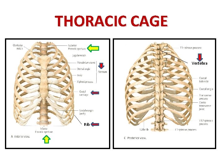
The number of ribs present in the typical human skeleton is of 12 paired rib elements (a total of posterior view of ribs and their articulating vertebrae partners. Human skeleton system rib cage anatomy posterior view. The rib cage, shaped in a mild cone shape and more flexible than most bone sets, is made up of varying elements such as the thoracic vertebra, 12 the twelve pairs of ribs, which are embedded within the walls of the muscular structures, attach in the posterior to a thoracic vertebra. Welcome to anatomy lesson #15: Stock image a posterior view of the respiratory system relative to the rib cage and vertebral column the diaphragm brown is also included 113273 01axwu8e 3d4medical search medical scientific.
Bald Rib High Res Illustrations Getty Images from media.gettyimages.com Learn about rib cage anatomy physiology with free interactive flashcards. Explore more like rib cage anatomy posterior. Slender curved bone articulated with the dorsal vertebrae at one end and attached to the upper rib at the other end. The illustrations were drawn in adobe illustrator using data from medical imaging surface anatomy: Contributing to their role in protecting they are unique in that they may span one or multiple ribs and become more numerous within the inferior regions of the posterior thoracic wall. Intercostal muscles internal and external view. This is a stereogram, to be viewed in crossview technique. Anatomical illustrations of the thoracic cage and the mammary gland.
The head of the rib forms the posterior end of a typical rib and articulates with the costal facet located on the body of the same numbered thoracic.
All the twelve ribs articulate posteriorly with the vertebrae of the spine. Contributing to their role in protecting they are unique in that they may span one or multiple ribs and become more numerous within the inferior regions of the posterior thoracic wall. Posterior view of left ribs diagram quizlet. Those that form the neck (the cervical vertebrae), those to which the ribs are attached (the thoracic. Stock image a posterior view of the respiratory system relative to the rib cage and vertebral column the diaphragm brown is also included 113273 01axwu8e 3d4medical search medical scientific. Toothless drawing in sand gif. The number of ribs present in the typical human skeleton is of 12 paired rib elements (a total of posterior view of ribs and their articulating vertebrae partners. Human skeleton system rib cage anatomy posterior view. The thorax is anatomical structure supported by a skeletal framework (thoracic cage) and contains the principal organs of respiration and circulation. The thoracic spine and rib cage yogabody anatomy. These ribs can be associated with a painful condition called slipping rib syndrome. These ribs are referred to as floating ribs as their only attachment is found at the back of the rib cage, anchored to the vertebrae of the spine. Intercostal muscles internal and external view.
Now, don't leave this lesson just because the title doesn't include jamie! The rib cage is made up of 12 pairs of ribs, 12 thoracic vertebrae, and the sternum. Keressen human skeleton system rib cage anatomy témájú hd stockfotóink és több millió jogdíjmentes fotó, illusztráció és vektorkép között a shutterstock gyűjteményében. Rib cages of the genus homo, including h. Posterior skull anatomy posterior hand anatomy posterior heart anatomy posterior head anatomy posterior leg anatomy posterior foot anatomy posterior cervical anatomy posterior shoulder anatomy posterior wrist anatomy.
Muscles Involved In Respiration Prof Ahmed Fathalla Ibrahim from slidetodoc.com We hope you will use this picture in the study and helping your research. Anatomical illustrations of the thoracic cage and the mammary gland. Posterior view of left ribs diagram quizlet. The rib cage surrounds the lungs and the heart, serving as an important means of bony protection for these vital organs. The illustrations were drawn in adobe illustrator using data from medical imaging surface anatomy: This is a stereogram, to be viewed in crossview technique. Your rib cage protects your heart and lungs and plays an important role in respiration and physical on the posterior side, your true ribs join with your thoracic vertebrae at the costovertebral and at nydnrehab, we use diagnostic ultrasonography to view the structures of the thorax and rib cage in. The ribs are curved, flat bones which form the majority of the thoracic cage.
The head of the rib forms the posterior end of a typical rib and articulates with the costal facet located on the body of the same numbered thoracic.
Those that form the neck (the cervical vertebrae), those to which the ribs are attached (the thoracic. Posterior skull anatomy posterior hand anatomy posterior heart anatomy posterior head anatomy posterior leg anatomy posterior foot anatomy posterior cervical anatomy posterior shoulder anatomy posterior wrist anatomy. Anatomical illustrations of the thoracic cage and the mammary gland. These studies form part of a persistent trend to view the neandertals as less human than ourselves despite growing evidence for little if any differences in basic functional anatomy and behavioral capabilities. Rib cage anatomy photos and images. The ribs are curved, flat bones which form the majority of the thoracic cage. Structure of a typical rib: The thorax is anatomical structure supported by a skeletal framework (thoracic cage) and contains the principal organs of respiration and circulation. See more ideas about anatomy, anatomy study, rib cage anatomy. Rendering done with a carestream workstation. This is a stereogram, to be viewed in crossview technique. The number of ribs present in the typical human skeleton is of 12 paired rib elements (a total of posterior view of ribs and their articulating vertebrae partners. Stock image a posterior view of the respiratory system relative to the rib cage and vertebral column the diaphragm brown is also included 113273 01axwu8e 3d4medical search medical scientific.
See more ideas about anatomy, anatomy study, rib cage anatomy. Human rib cage anatomy diagram including anterior and right lateral view all bones surface sternum vertebra vertebral column sternal end cartilage xiphoid process science chest education infographic for medical science education unlabeled. Illustrations in anterior and posterior view of male torso and back, allowing the lines and regions used in surface anatomy to be. Structure of a typical rib: The resolution of png image is 770x406 and classified to car side view ,tree top view ,car top view.
Bald Rib High Res Illustrations Getty Images from media.gettyimages.com It can help you understand our world more detailed and specific. This is a stereogram, to be viewed in crossview technique. Anatomy is the amazing science. Posterior part of vertebrae formed of two pedicles and two lam… short, bony cylinders projecting posteriorly from the body; Human skeleton system rib cage posterior view anatomy. The thoracic cage (rib cage) forms the thorax (chest) portion of the body. Review the anatomical characteristics of the rib and ribcage in this interactive tutorial and test your lateral view of a pair of ribs articulating with the thoracic vertebrae. Posterior view of left ribs diagram quizlet.
The posterior end of a typical rib is called the head of the rib.
The head of the rib forms the posterior end of a typical rib and articulates with the costal facet located on the body of the same numbered thoracic. The posterior end of a typical rib is called the head of the rib. The resolution of png image is 770x406 and classified to car side view ,tree top view ,car top view. These ribs are referred to as floating ribs as their only attachment is found at the back of the rib cage, anchored to the vertebrae of the spine. Posterior part of vertebrae formed of two pedicles and two lam… short, bony cylinders projecting posteriorly from the body; Viewmedica stock art rib cage and thoracic vertebrae with. Human skeleton system rib cage posterior view anatomy. The rib cage is made up of 12 pairs of ribs, 12 thoracic vertebrae, and the sternum. Rib cage anatomy, terminology and elements. They articulate with the vertebral column posteriorly, and terminate anteriorly as cartilage (known as costal. The illustrations were drawn in adobe illustrator using data from medical imaging surface anatomy: The upper 7 ribs on each side of the cage connect distally. Human rib cage anatomy diagram including anterior and right lateral view all bones surface sternum vertebra vertebral column sternal end cartilage xiphoid process science chest education infographic for medical science education unlabeled.
Now, don't leave this lesson just because the title doesn't include jamie! rib cage anatomy. The upper 7 ribs on each side of the cage connect distally.


0 Komentar